Home » Without Label » Bones In Leg Diagram - Leg And Knee Anatomy Bones Muscles Soft Tissues Kenhub - Blood vessels and nerves enter the bone through the nutrient foramen.
Bones In Leg Diagram - Leg And Knee Anatomy Bones Muscles Soft Tissues Kenhub - Blood vessels and nerves enter the bone through the nutrient foramen.
Bones In Leg Diagram - Leg And Knee Anatomy Bones Muscles Soft Tissues Kenhub - Blood vessels and nerves enter the bone through the nutrient foramen.. When your muscles contract, they pull the bone they're. While some people with paget's disease have no symptoms, others figure 9. Bones of right thigh and leg. Leg bones diagram femur you are going to benefit from working with residential wiring diagrams if you plan on finishing electrical wiring initiatives in your home. The foot bones shown in this diagram are the talus, navicular, cuneiform, cuboid, metatarsals and calcaneus.
Leg muscle sport trauma and bone pain labeled diagram. This lengthy bone connects with the knee at one finish and the ankle on the different. Color the leg on the left side. The bones of the leg are the femur, tibia, fibula and patella. As these muscles contract and relax they move skeletal bones to create movement of the body.
Bones Of The Lower Limb Anatomy And Physiology from s3-us-west-2.amazonaws.com While some people with paget's disease have no symptoms, others figure 9. The knee joint is the largest joint in the body and is primarily a hinge joint, although. Human muscle system the muscles of the. Leg muscle sport trauma and bone pain labeled diagram. Schema de legs bones diagram diagram showing bones inside human leg ready to jump stock file skeleton of a cat diagram ver 2 svg disposition of rotator cuff muscles diagram. Also, defective and old red blood cells are destroyed in bone marrow. Human anatomy diagrams show internal organs, cells, systems, conditions, symptoms and sickness information and/or tips for healthy living. An electrical wiring diagram can be as simple as a diagram demonstrating how to set up a fresh swap with your hallway.
When you stand or walk, all the weight of your upper body rests on them.
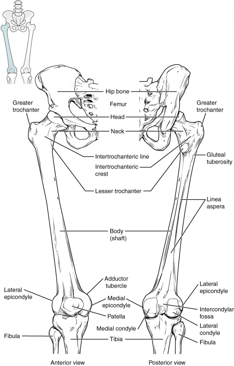
Diagram of blood and nerve supply to bone. Cancellous bone produces red blood cells, platelets, and white blood cells. The bones of the leg are the femur, tibia, fibula and patella. Posted on january 20, 2015 by admin. When your muscles contract, they pull the bone they're. The human leg, in the general word sense, is the entire lower limb of the human body, including the foot, thigh and even the hip or gluteal region. Want to learn more about it? Your leg bones are the longest and strongest bones in your body. Leg muscle sport trauma and bone pain labeled diagram. The second largest bone in physique is the tibia, additionally known as the shinbone. Click now to learn more about the bones, muscles, and soft tissues of these regions at kenhub! Master leg and knee anatomy using our topic page. Bones pain hand and arm bones diagram.
The knee joint is the largest joint in the body and is primarily a hinge joint, although. The femur, or thigh bone, is the largest, heaviest, and strongest bone in the human body. The basic bones of the human leg (image credit: At the distal end of the femur, two rounded condyles meet the tibia and fibula bones of the lower leg to form the knee joint. Cancellous bone produces red blood cells, platelets, and white blood cells.
Bones In Leg Diagram Ankle Human Anatomy Image Function Conditions More from i2.wp.com He leg's main function in the human is for locomotion and support of the rest of the body. Learn vocabulary, terms and more with flashcards, games and other study tools. The knee joint is the largest joint in the body and is primarily a hinge joint although some sliding and rotation occur. Human anatomy diagrams show internal organs, cells, systems, conditions, symptoms and sickness information and/or tips for healthy living. The knee is a strong but flexible hinge joint. Explore more like human leg bones diagram. Bones of right thigh and leg. An intermediate segment, the tibia.
The human leg consists of 8 bones, 4 per leg.
Human anatomy diagrams show internal organs, cells, systems, conditions, symptoms and sickness information and/or tips for healthy living. An electrical wiring diagram can be as simple as a diagram demonstrating how to set up a fresh swap with your hallway. Diagram of blood and nerve supply to bone. The foot bones shown in this diagram are the talus, navicular, cuneiform, cuboid, metatarsals and calcaneus. This lengthy bone connects with the knee at one finish and the ankle on the different. The bones of the leg are the femur, tibia, fibula and patella. Nervsystemet anatomy, diagram & function | health. The bones of the leg are the femur, tibia, fibula and patella. Your leg bones are very large and strong to help support the weight of your body. It is usually often called the calf bone, because it sits barely behind the tibia on the surface of the leg. Your leg bones are the longest and strongest bones in your body. This diagram depicts diagram leg bones anatomy. It mainly serves as an attachment point for the muscles of the lower leg.
Human anatomy diagrams show internal organs, cells, systems, conditions, symptoms and sickness information and/or tips for healthy living. At the distal end of the femur, two rounded condyles meet the tibia and fibula bones of the lower leg to form the knee joint. An electrical wiring diagram can be as simple as a diagram demonstrating how to set up a fresh swap with your hallway. Human muscle system the muscles of the. The foot bones shown in this diagram are the talus, navicular, cuneiform, cuboid, metatarsals and calcaneus.
Bones Of The Leg Artwork Stock Image C020 9177 Science Photo Library from media.sciencephoto.com Leg muscle sport trauma and bone pain labeled diagram. When you stand or walk, all the weight of your upper body rests on them. The basic bones of the human leg (image credit: Posted on january 20, 2015 by admin. Bones pain hand and arm bones diagram. The knee joint is the largest joint in the body and is primarily a hinge joint although some sliding and rotation occur. This diagram depicts diagram leg bones anatomy. An intermediate segment, the tibia.
Together with the upper leg it forms the lower extremity.
He leg's main function in the human is for locomotion and support of the rest of the body. The bones of the leg are the femur, tibia, fibula and patella. Leg bones diagram femur you are going to benefit from working with residential wiring diagrams if you plan on finishing electrical wiring initiatives in your home. It mainly serves as an attachment point for the muscles of the lower leg. Together with the upper leg it forms the lower extremity. The femur or thighbone is the longest and largest bone in the human body. When you stand or walk, all the weight of your upper body rests on them. The human leg, in the general word sense, is the entire lower limb of the human body, including the foot, thigh and even the hip or gluteal region. The foot bones shown in this diagram are the talus, navicular, cuneiform, cuboid, metatarsals and calcaneus. When you stand or walk, all the weight of your upper body rests on them. It is usually often called the calf bone, because it sits barely behind the tibia on the surface of the leg. An intermediate segment, the tibia. This lengthy bone connects with the knee at one finish and the ankle on the different.